
Key messages
- Diagnosis of plantar heel pain can be made from a clinical assessment (Chimutengwende-Gordon, O’Donnell & Singh, 2010, Martin et al, 2014)
- Findings from imaging seldom change the management approach for people with plantar heel pain (Chimutengwende-Gordon, O’Donnell & Singh, 2010)
- Another common cause of pain beneath the heel is fat pad contusion (Landorf, Simons, Jordan & Rathleff, 2017)
- Baxter’s neuropathy is often missed as a diagnosis (Landorf, Simons, Jordan & Rathleff, 2017)
- A patient may have more than one condition occurring simultaneously (Chimutengwende-Gordon, O’Donnell & Singh, 2010)
- The impact of non-biological factors and how these contribute to the pain experience should not be overlooked
- Imaging can support a clinical diagnosis but is not recommended as standard practice (Landorf, Simons, Jordan & Rathleff, 2017; Riel, Cotchett, Delahunt, Rathleff, Vicenzino, Weir & Landorf, 2017)
- Differential diagnosis and imaging should be considered when there is persistent pain (despite appropriate treatment), or atypical pain (Chimutengwende-Gordon, O’Donnell & Singh, 2010)
Differential diagnosis
Structures under the heel include skin, nerves, blood vessels, subcalcaneal bursa, plantar fascia, fat pad and the bony calcaneus. Differential diagnoses of pain beneath the heel are numerous and examples include, but are not limited to, fat pad contusion, Baxter’s nerve entrapment, plantar fascia rupture, plantar fibroma, L5-S1 radiculopathies, calcaneal stress fracture, retrocalcaneal bursitis, calcaneal apophysitis (Sever’s) in adolescents, soft tissue or bony tumours, deep vein thrombosis, complex regional pain syndrome type 1, bone or tissue infections, rheumatoid arthritis, gout and fibromyalgia (Landorf, Simons, Jordan & Rathleff, 2017; personal communication M Cotchett, October 31 2019; Thomas et al, 2010; Chimutengwende-Gordon, O’Donnell & Singh, 2010).
However, as previously mentioned, a thorough history and objective examination is generally adequate to diagnose plantar heel pain.
When considering pain of purely biological origins, heel pain can be classified into the following five categories:
- Mechanical (most common)
- Neurological
- Arthritic
- Traumatic
- Other systemic (Thomas et al, 2010)

Mechanical causes of plantar heel pain refer to issues around loading, whether that be due to overweight or obesity, repetitive stress or biomechanical factors (Thomas et al, 2010). Neurological causes of heel pain arise due to proximal and/or distal nerve impingement or irritation (Thomas et al., 2010). Traumatic causes include problems such as calcaneal stress fractures and fat pad contusions which generally occur from a mechanism of injury such as a fall from height.
Another source of traumatic injury to the heel resulting in heel pain, apart from fat pad bruising, is calcaneal stress fracture. This can present in active individuals where weight bearing activities, such as running, aggravate their pain. Onset is insidious, with tenderness on palpation on the lateral or medial calcaneus. Pain may be reproducible by squeezing the calcaneus on both sides (paying attention to the bony aspect of the calcaneus, as opposed to the soft tissue structures on the plantar surface of the heel) (Landorf, Simons, Jordan & Rathleff, 2017; M. Cotchett personal communication October 31 2019).
Neurological causes
Be suspicious of neurological causes if there is:
- a history of surgery or trauma to the heel
- altered sensation in the foot
- pain which commences in the lower back and radiates down the lower limb
In these cases, it is important to rule out heel pain due to systemic disease such as diabetic neuropathies, alcoholism or nutritional inadequacies – particularly if the pain is bilateral (Thomas et al., 2010).
Aside from a thorough subjective examination, further objective tests may provide clues in diagnosing the differing sources of pain beneath the heel. For example, if a radiculopathy is suspected, neurodynamic testing, such as a slump test or straight leg raise, can confirm involvement from more proximal structures. Another probable diagnosis is nerve entrapment. Entrapment is common in the tarsal tunnel due to the proximity of nerves, blood vessels and osteofibrous structures in a small space (Moroni, Zwierzina, Starke, Moriggl, Montesi & Konschake, 2019), as well as the potential for space-occupying lesions (such as varicosities, ganglia) to be present, narrowing the space even further.
Baxter’s neuropathy is the most common cause of neurological heel pain, and is often overlooked (Moroni, Zwierzina, Starke, Moriggl, Montesi & Konschake, 2019; Landorf, Simons, Jordan & Rathleff, 2017). Clinically, people with a lateral plantar nerve entrapment will present with non-specific pain in the medial aspect of the heel and medial ankle region, with minimal complaints of pins and needles or numbness (Landorf, Simons, Jordan & Rathleff, 2017, M. Cotchett personal communication October 31 2019). It can also present in combination with plantar heel pain, in around 50% of patients (Landorf, Simons, Jordan & Rathleff, 2017). A way to assess for possible Baxter’s nerve entrapment is by asking the patient to abduct their little toe, and compare sides, as Baxter’s nerve innervates the abductor digiti minimi muscle. A similar assessment can be conducted with the abductor hallucis muscle, which abducts the big toe, to check for medial plantar nerve entrapment (M. Cotchett personal communication October 31 2019).
A 2019 study (Moroni, Zwierzina, Starke, Moriggl, Montesi & Konschake) investigated topographical sites of nerves in the tarsal tunnel and areas of possible nerve entrapment. They found that in approximately 80% of cases potential entrapment sites for the following nerves were found within a 5mm radius of each other; medial plantar nerve, lateral plantar nerve, Baxter’s nerve (first branch, lateral plantar n.) and medial calcaneal branch of tibial nerve. See image below.

Clinically, this ‘circle method’ or 5mm radius, can give the clinician confidence that nerve entrapment in the tarsal tunnel follows a regular pattern in 4 out of 5 cases. Results contrast to previous studies stating that entrapment of Baxter’s nerve occurs either proximally at the medial calcaneal tuberosity (where the plantar fascia originates), or distally between the quadratus plantae muscle and abductor hallucis fascia. In any case, this appears quite similar in practice to the Triple Compression Stress Test and Tinel’s sign for tarsal tunnel nerve entrapment.
Although some of the tests listed may have high specificity or sensitivity, remember that these are not necessarily useful indicators of diagnostic accuracy and rarely does one test singly prove or refute a diagnosis (Herbert, Jamvedt, Hagen & Mead, 2011). In addition, it is not uncommon for people to present with more than one condition at the same time (Chimutengwende-Gordon, O’Donnell & Singh, 2010).
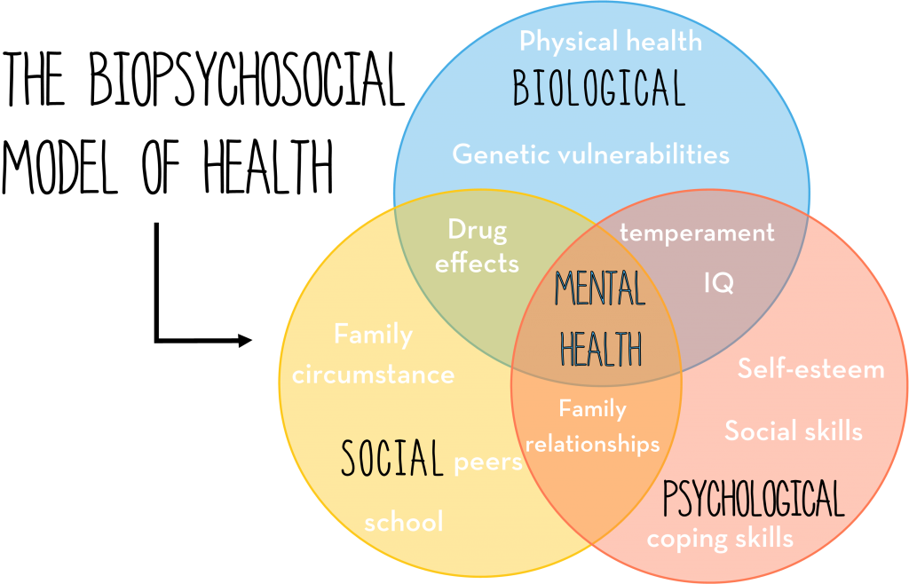
Biopsychosocial model of health
Finally, whilst categorising causes of heel pain into discrete boxes helps to simplify understanding, how people experience pain is complex. Consider how factors which lie outside of the individual, or the social determinants of health, influence a person’s lived experience. Housing, employment, education, access to transport, income – all play a part in determining individual health outcomes, for better or worse.
People with heel pain are more likely to experience depression, anxiety and stress (Cotchett, Munteanu & Landorf, 2016) and the elements which give rise to someone feeling depressed, anxious or stressed are often multi-factorial and complex. Researchers in 2017 (Cotchett, Lennecke, Medica, Whittaker & Bonnano) studied the relationship between catastrophising and kinesiophobia on pain and function in people with plantar heel pain. They found a significant association between foot function and fear of movement and pain catastrophising, and first step pain and pain catastrophising. Outcome measures such as the Pain Catastrophising Scale can be used to objectively assess the impact of psychological factors in a person’s presentation. Although causality has not been established, clinician’s managing people with plantar heel pain should consider the impact of kinesiophobia and catastrophising on pain and function (Cotchett, Lennecke, Medica, Whittaker & Bonnano, 2017).
In other words, the patient should always be considered in the context of broader psychosocial factors – a biopsychosocial approach – and not just their physiology.
Imaging
Imaging may help to confirm a clinical diagnosis of plantar heel pain but is not recommended as standard practice (Landorf, Simons, Jordan & Rathleff, 2017; Riel, Cotchett, Delahunt, Rathleff, Vicenzino, Weir & Landorf, 2017). As with other conditions, imaging findings must correlate with clinical symptoms and there are instances where asymptomatic people have abnormal imaging findings, and where symptomatic people have normal imaging findings.
When further investigation into heel pain is warranted, Magnetic Resonance Imaging (MRI), x-ray and ultrasound are all potential imaging modalities. Ultrasound can detect thickness of the plantar fascia, hypoechoic characteristics of the tissue and plantar fibroma (fibrous tissue mass), of which the former two are frequently found in people with plantar heel pain (Chimutengwende-Gordon, O’Donnell & Singh, 2010). Plain radiographs can be used to detect suspected fractures, calcaneal heel spurs and may highlight other bony changes such as osteoarthritis. MRI highlights in greater detail bone, soft-tissue, vascular and neural structures and can detect infection, stress fractures, nerve entrapments and fascia rupture, among others (Chimutengwende-Gordon, O’Donnell & Singh, 2010).
A 2010 retrospective study on MRI in plantar heel pain revealed (Chimutengwende-Gordon, O’Donnell & Singh, 2010):
In fifty patients with persistent pain (despite appropriate treatments of at least 3 months)
- Findings were normal in 14% of cases, despite the presence of clinical symptoms
- Diagnosis was confirmed in 76%
- Almost 1 in 5 of the above 76% had another condition simultaneously (such as partial plantar fascia tear, Achilles’ tendinopathy)
- Only 2% resulted in a diagnosis which changed clinical management (posterior ankle impingement)
Patients were classified as having atypical pain if they experienced any of the following, irrespective of symptom duration:
- night pain
- acute onset or worsening of pain
- tenderness or pain at the lateral or medial heel
- pain posterior to the plantar fascia insertion
- sizeable swelling
- neurological symptoms
Imaging findings from the above cohort revealed a significant correlation between:
- night pain and calcaneal bone marrow oedema
- acute pain and plantar fascia tear
Clinical findings in the atypical patient presentation which did not relate to plantar fascia pathology included: osteoarthritis, osteoporosis, calcaneal stress fracture, tarsal tunnel ganglion, tibialis posterior tendinopathy, retrocalcaneal bursitis, inflamed abductor hallucis and a plantar arterio-venous malformation.
Similar results were found in a 1999 study (Grasel, Schweitzer, Kovalovich, Karasick, Wapner, Hecht & Wander) with perifascial oedema the most common MRI finding, followed by calcaneal bone marrow abnormalities (oedema, lesions), fascial tear, history of trauma and fascia thickening. A key finding of interest from this study is that bone bruising, or oedema, in plantar heel pain behaves differently to bony bruising elsewhere in the body – it does not respond typically and heal within a few weeks. However, another key finding was that there was no difference to patient outcomes between those with or without bone marrow oedema. Again, this supports the notion that it is rare MRI findings change management of plantar heel pain.
Prognosis
- General rule for prognosis: the longer symptoms have been present, the longer they will take to resolve (Thomas et al, 53 54)
- Females and those with bilateral heel pain have a less favourable prognosis (Hansen, Krogh, Ellingsen, Bolvig & Fredburg, 2018)
- Prognosis does not appear to be affected by plantar fascia thickness or heel spur (Hansen, Krogh, Ellingsen, Bolvig & Fredburg, 2018).
A 2018 Danish (Hansen, Krogh, Ellingsen, Bolvig & Fredburg) longitudinal study found:
- Only 1 in 5 people are symptom free after one year
- After 10 years, almost half still had symptoms (even though most symptoms were minimal)
- In people who are symptom free after 10 years, the average duration of symptoms is 2 years
However, participants included in the study tended to have more severe cases of heel pain.
Sky Daly-Holt
Bachelor of Applied Science and Master of Physiotherapy Practice 4th year studentDecember 2019